Note the gender difference in distance between both cirsta iliaca anterior superior (distantia interspinosa), the. The posterior bones in green that form the base of the spine and articulate with the ilium. Pelvic floor by amrit kaur 19360 views. Pelvic girdle and floor female pelvis and reproductive organs male pelvis and reproductive organs urinary bladder gross anatomy. Plane of mid cavity (plane of greatest pelvic dimensions).
Pelvic Bone Labeling - Human Anatomy Body from www.anatomylibrary99.com Pelvic surgery requires a comprehensive knowledge of the pelvic anatomy to safely attain access, maximize exposure, ensure hemostasis, and avoid injury to viscera, blood vessels, and nerves. Anatomical points for obstetric analgesia. Abbreviations used in figures 1 through 4: Related online courses on physioplus. Postoperativ view of abdomen after tep operation by anpol42. The geometry of bony pelvis front view of the male and female pelvis. It can help you understand our world more detailed and specific. Anatomyzone is youtube's most highly subscribed anatomy channel, with video tutorials on all areas of anatomy.
This anatomy section promotes the use of the terminologia anatomica, the international standard of anatomical nomenclature.
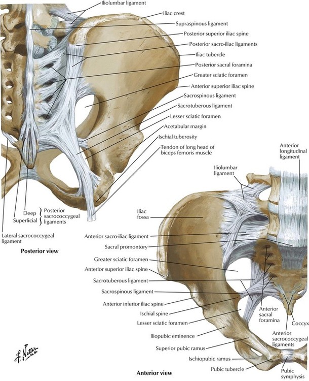
Some put posterior and downward force on the ilium to pull. Pelvic floor by amrit kaur 19360 views. Anatomynote.com found pelvic region posterior view from plenty of anatomical pictures on the internet. Contemporary views on female pelvic anatomy. Female pelvic bone anatomy bony pelvis anatomy pelvic girdle side view pelvic bone landmarks front view male pelvis anatomy pelvic girdle anatomy lateral view anatomy of pelvic area and hip sacroiliac joint posterior view right hip bone medial view hip bone structure anatomy. Pelvic skeleton includes two hip bones, sacrum and coccyx. But understanding this level of skeletal anatomy will make it easier to understand muscle and how can you quickly visually assess a client's propensity towards a pelvic tilt? This anatomy section promotes the use of the terminologia anatomica, the international standard of anatomical nomenclature. Female pelvis ppt by mayil rasamani 152255 views. # denotes the levator ani attachment to room analogy; We hope you will use this picture in the study and. The sacrum and coccyx forms part of the pelvis and the pelvis attaches to the spine at the runs from the posterior pelvis and attaches to the asis to the ribs and a sheath over the transverse abdominus. A thorough understanding of pelvic anatomy is essential for clinical practice.
A thorough understanding of pelvic anatomy is essential for clinical practice. The line of attachment of the pubocervical fascia to the levator ani is arcus tendineus fascia pelvis. Vides a discussion of the contemporary understanding. Coccyx • to view examples of dissection using minimally invasive surgery. Pass between the middle of the posterior surface of the symphysis pubis and the junction between.
5: Pelvis and Perineum | Basicmedical Key from basicmedicalkey.com Atfp, arcus tendineus fasciae after the viscera of the abdomen and pelvis have been removed from a cadaver the general shape and contour of the posterior abdominal wall may be. Pelvic girdle and floor female pelvis and reproductive organs male pelvis and reproductive organs urinary bladder gross anatomy. Female pelvis ppt by mayil rasamani 152255 views. # denotes the levator ani attachment to room analogy; What is the collateral whiteside jl, et al. The bony pelvis & gender differences in pelvic anatomy. Anatomyzone is youtube's most highly subscribed anatomy channel, with video tutorials on all areas of anatomy. The geometry of bony pelvis front view of the male and female pelvis.
Pass between the middle of the posterior surface of the symphysis pubis and the junction between.
The line of attachment of the pubocervical fascia to the levator ani is arcus tendineus fascia pelvis. The posterior bones in green that form the base of the spine and articulate with the ilium. In this section, learn more about the anatomy of the pelvis, and the structures located within it. Anatomynote.com found pelvic region posterior view from plenty of anatomical pictures on the internet. Pelvic surgery requires a comprehensive knowledge of the pelvic anatomy to safely attain access, maximize exposure, ensure hemostasis, and avoid injury to viscera, blood vessels, and nerves. Anterior to obturator canal insertion: Anatomical points for obstetric analgesia. The posterior abdominal wall is a musculoskeletal structure formed by the posterior abdominal muscles, their fascia, the lumbar vertebrae and the pelvic girdle. Abdominal and pelvic anatomy encompasses the anatomy of all structures of the abdominal and pelvic cavities. Anatomy of pelvis & perineum by profgoodnewszion 74013 views. But understanding this level of skeletal anatomy will make it easier to understand muscle and how can you quickly visually assess a client's propensity towards a pelvic tilt? ƒ iliolumbar ƒ lateral sacral ƒ superior gluteal. ƒ organs and structures of the female pelvis.
Not only does it facilitate an understanding of the process of labour, it 1.4the blood supply of the uterus, fallopian tube and ovary (posterior view). Related online courses on physioplus. We think this is the most useful anatomy anatomy is the amazing science. Some put posterior and downward force on the ilium to pull. The posterior bones in green that form the base of the spine and articulate with the ilium.
Anatomyzone is youtube's most highly subscribed anatomy channel, with video tutorials on all areas of anatomy.
Organs and the anococcygeal raphe. From the tip of the sacral promontory to the upper border of the symphysis pubis. It can help you understand our world more detailed and specific. The posterior abdominal wall is a musculoskeletal structure formed by the posterior abdominal muscles, their fascia, the lumbar vertebrae and the pelvic girdle. Note the gender difference in distance between both cirsta iliaca anterior superior (distantia interspinosa), the. ƒ iliolumbar ƒ lateral sacral ƒ superior gluteal. This is formed by the fusion of the ilium, ischium and pubis. Safe access to retroperitoneal structures. A thorough understanding of pelvic anatomy is essential for clinical practice. Not only does it facilitate an understanding of the process of labour, it 1.4the blood supply of the uterus, fallopian tube and ovary (posterior view). Female pelvis ppt by mayil rasamani 152255 views. Pelvic surgery requires a comprehensive knowledge of the pelvic anatomy to safely attain access, maximize exposure, ensure hemostasis, and avoid injury to viscera, blood vessels, and nerves. Gluteal muscles are located posteriorly and are closely associated with the ilium.
The posterior bones in green that form the base of the spine and articulate with the ilium pelvic anatomy. Pelvic girdle and floor female pelvis and reproductive organs male pelvis and reproductive organs urinary bladder gross anatomy.


0 Comments